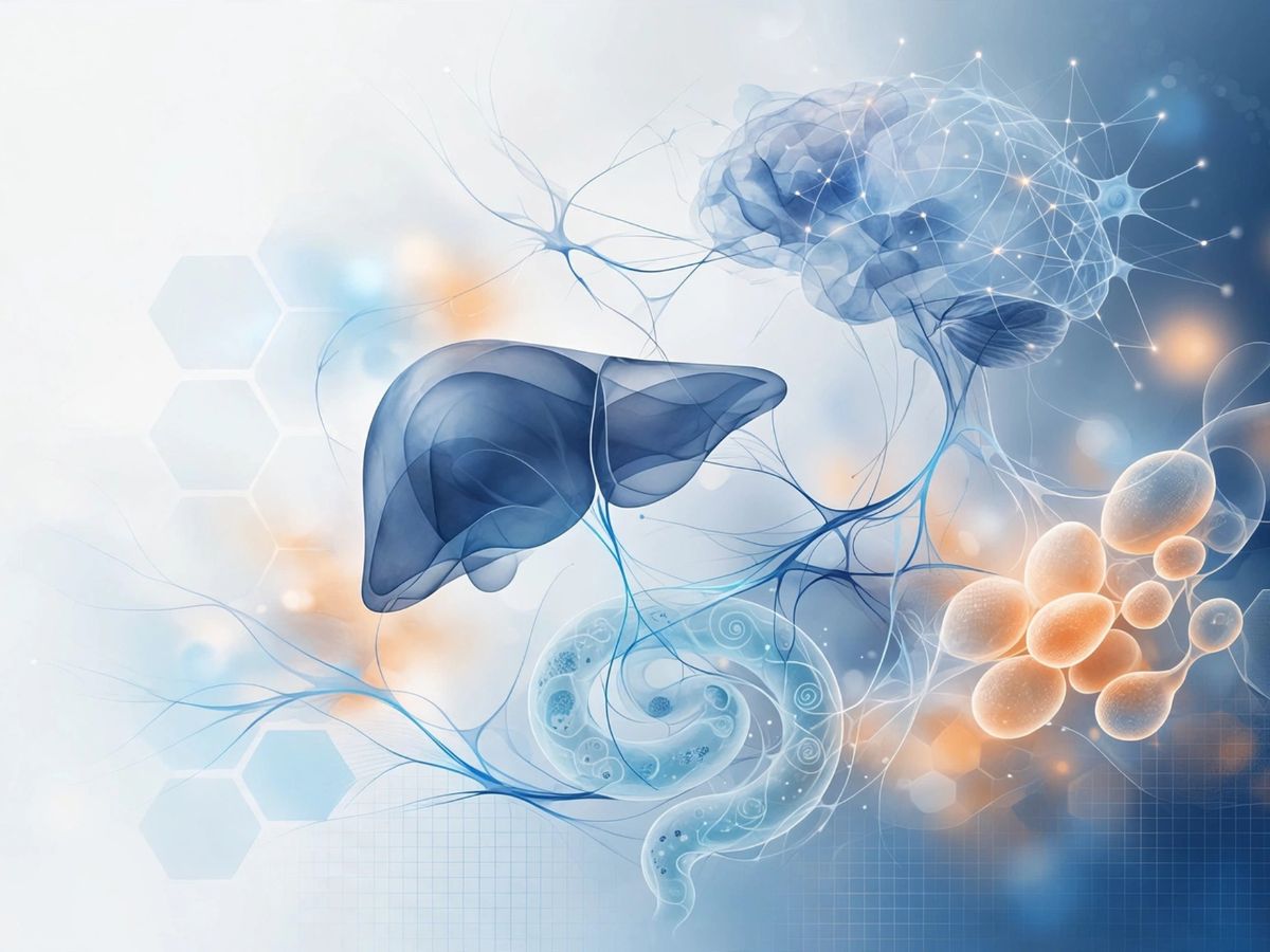
Spatial Mass Spectrometry
Target engagement and proof of mechanism are no longer optional in early drug development. Regulators and investors expect tissue-level evidence to support those claims, yet most preclinical packages cannot deliver it. Our mass spectrometry imaging (MSI) platform maps where a drug goes, what it does, and what processes it affects. These measurements are performed directly on intact tissue sections, without the need for extraction or labelling. MSI can also provide new insights into disease development on a molecular level.
Why Mass Spectrometry imaging matters in drug development
MSI is increasingly essential in drug development, as traditional approaches do not always reveal what truly happens inside tissues. For decades, preclinical pharmacokinetic studies have relied on plasma concentrations as a proxy for drug exposure. However, plasma levels often fail to reflect how a compound distributes within target organs and tissues.
A drug may show excellent plasma exposure while failing to reach the intended tissue, accumulating in off-target regions, or distributing unevenly within an organ. It may even reach the correct tissue, but not the specific cell population responsible for disease. These critical aspects of drug behavior cannot be captured through plasma measurements alone and only become visible when studying the tissue directly.
Conventional tissue analyses each carry significant limitations. Whole-body autoradiography requires radiolabeled compounds and lacks molecular specificity. Liquid Chromatography-Tandem Mass Spectrometry (LC-MS/MS) of tissue homogenates deliver accurate concentration data but eliminate all spatial information. Immunohistochemistry provides spatial context but depends on specific antibodies and does not directly measure the drug or its metabolites. As a result, these techniques cannot fully link molecular information to tissue structure and function.
Mass spectrometry imaging overcomes these limitations by enabling the direct measurement and visualization of molecules on intact tissue sections, without the need for labelling. It provides spatially resolved, compound-specific information, allowing researchers to see exactly where drugs and metabolites are located within tissues and how it interacts with other biomolecular species.
Importantly, this molecular data can be directly combined with histological information, offering a comprehensive view of drug distribution in the context of tissue architecture. By delivering both spatial and molecular insight, mass spectrometry imaging helps answer key questions in pharmaceutical research. It allows researchers to determine whether a drug reaches its intended target, identify potential off-target accumulation, and better understand how distribution relates to efficacy and toxicity. In doing so, it supports more informed decision-making and de-risks drug development at an early stage.
What is Mass Spectrometry imaging?
Mass spectrometry imaging (MSI) is an analytical technique that reveals the spatial distribution of molecules within thin tissue sections. During the measurement, an ionization probe scans across the tissue surface, generating molecular ions that are measured based on their mass-to-charge ratio. At every position on the tissue, a full mass spectrum is acquired, meaning each pixel in the resulting image contains a detailed molecular profile of that specific location.
From a single tissue section, MSI can map the localization of a drug and its metabolites alongside endogenous molecules such as lipids, small metabolites, peptides, and even proteins, that reflect the biochemical state of the tissue. Importantly, this is achieved without the need for extraction or labelling. Compounds are detected directly based on their molecular mass, enabling clear differentiation between the parent drug and structurally related metabolites within a single experiment.
By combining spatial precision with molecular specificity, MSI provides unique insight into how compounds interact with biological systems at the tissue level.
How does it work?
The MSI analysis is performed using matrix-assisted laser desorption/ionization (MALDI). A finely controlled laser scans across the tissue surface, releasing and ionizing molecules at each position. At every point, a full molecular spectrum is recorded, allowing the reconstruction of detailed distribution of maps that show exactly where compounds are located within the tissue.
Our platform is based on a high-resolution TimsTOF instrument, which adds an additional dimension of separation. This enhances molecular specificity and helps distinguish closely related compounds, such as drug metabolites or structurally similar lipids, within complex biological samples.
MSI data are processed to generate ion distribution images and, where quantitative analysis is required, to create tissue concentration maps calibrated using reference standards. To measure absolute drug concentrations at tissue level, TNO offers the possibility to use microtracer labeled compounds.
By (spatial) mass spectrometry data, paired with AMS-based microtracer quantification, absolute drug metabolite concentrations can be assigned at the tissue level, with the spatial distribution pattern providing the mechanistic context that bulk tissue LC-MS/MS measurement cannot. The resulting dataset combines absolute quantification with spatial resolution in a single analytical workflow.
Because MSI is performed on the same tissue section from which histological stains and immunofluorescence can be obtained, the molecular distribution maps can be co-registered with morphological tissue context. This overlay directly correlates drug distribution with histological lesion boundaries, disease-relevant cell populations, or biomarker expression patterns.
Interpretation is strengthened through collaboration with our in-house pathologists and domain specialists, who help place the spatial molecular data in the right biological and disease context. Together, this provides the mechanistic interpretation layer that transforms distribution data into proof-of-mechanism evidence.
What mass spectrometry imaging delivers that conventional methods cannot
Drug and metabolite distribution at the sub-organ compartment to cellular level, without extraction or labelling. Simultaneous mapping of parent compounds and metabolites in a single analytical run. Direct spatial correlation between drug distribution and histological or molecular tissue features.
Detection of off-target tissue accumulation or target compartment exclusion that plasma PK cannot reveal. Combined with AMS microtracer quantification: absolute tissue concentration combined with spatial resolution.
Applications across indication areas
In atherosclerosis programmes, the therapeutic target is typically a specific cell population within a complex, spatially heterogeneous lesion. Whether a compound reaches the macrophage-rich necrotic core, the fibrous cap, or the medial smooth muscle layer is mechanistically decisive, and invisible to plasma pharmacokinetics.
MSI maps drug distribution within aortic root cross-sections, directly correlated with Oil Red O staining or immunofluorescence marking of plaque subpopulations, providing the spatial proof of target site penetration that regulatory and partnering audiences increasingly require.
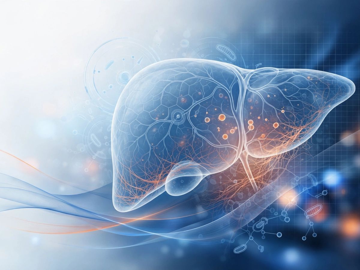
In MASLD and MASH programmes, hepatic drug distribution varies across the lobular architecture. Portal zone hepatocytes, centrilobular regions and activated stellate cell populations may respond differently to the same nominal tissue exposure. MSI resolves compound distribution at lobular resolution and can simultaneously map endogenous lipid species and metabolic markers that confirm target pathway engagement.
Blood-brain barrier penetration is one of the most consequential and frequently undercharacterised properties in CNS drug development. Plasma exposure provides no information on regional brain distribution, and whole brain homogenate data misses the spatial heterogeneity that determines whether therapeutic concentrations are reached in the relevant structure.
MSI maps compound distribution across brain regions including cortex, hippocampus, striatum and cerebellum in the same section, distinguishing grey and white matter penetration and identifying any regional exclusion that would compromise target engagement.
TNO's role: integrated spatial evidence within a single workflow
We integrate MSI within the same translational research environment that generates the tissue. The disease model, the tissue, the spatial distribution data, the histopathological analysis, and, where relevant, the AMS-based metabolic flux and microtracer quantification data are all produced within a single coordinated workflow. This integration is what allows the data to be interpreted as a coherent mechanistic story rather than a collection of separately commissioned measurements.
Spatial mass spectrometry data are interpretable only in the context of the diseased tissue they describe. Our platform is applied within validated, well-characterised disease models including the TNO Ldlr-/-.Leiden mouse for MASLD and atherosclerosis, the APOE*3-Leiden and APOE*3-Leiden.huCETP models for cardiovascular indications, renal models and fibrosis models covering skin, lung, liver and kidney. The tissue phenotypes in these models are extensively characterized.
For biotech and pharma teams building the mechanistic case for an IND filing, a partnering discussion or a preclinical publication, the combination of tissue-level distribution data, proof of target site penetration, and pathway-level mechanistic readouts represents the kind of evidence package that advances a programme's credibility at a stage where credibility is still being established.
We build that package within a single integrated study, rather than requiring multiple contracted studies across multiple providers with the coordination overhead and data integration risk that entails.
Platform integration: spatial mass spectrometry alongside AMS and HistoSuite
The three analytical platforms described across this series, AMS-based metabolic flux analysis, HistoSuite AI histopathology, and spatial mass spectrometry, are designed to be applied within the same preclinical study on the same tissue material.
From a single efficacy study with tissue collection: spatial drug distribution maps (this platform), quantitative histopathological lesion readouts (HistoSuite), and metabolic pathway flux data (AMS microtracer). Together, these constitute the mechanistic evidence layer that sits between biomarker readouts and clinical proof of concept.
Work with us
If you are building the mechanistic evidence package for your programme, we would welcome the opportunity to be part of that conversation. Whether the priority is demonstrating target site penetration, resolving an unexpected pharmacokinetic finding, or strengthening the mechanistic narrative ahead of an IND filing or partnering discussion, our team can outline the study design and analytical approach most relevant to your compound and indication.
Contact us
-

Robert Ostendorf
Functie:Senior Business Development Manager translational pharmaceutical researchI am passionate about TNO’s unique expertise in translational (preclinical) research. My work focuses on efficacy, ADME/DMPK and target safety of new pharmaceutical interventions, with special interest in cardiovascular, metabolic and fibrotic diseases, and biomarker research.
Looking for contract research support, or see opportunities for collaboration? Please, reach out.
-
Standplaats:Leiden - Sylviusweg
-
Email:Email Robert
-
LinkedIn:Robert on LinkedIn
-
Get inspired
14C-Pulse-Chase Technology

Obesity
Diabetic kidney disease

MASH and liver fibrosis
HistoSuite: AI-powered histopathology